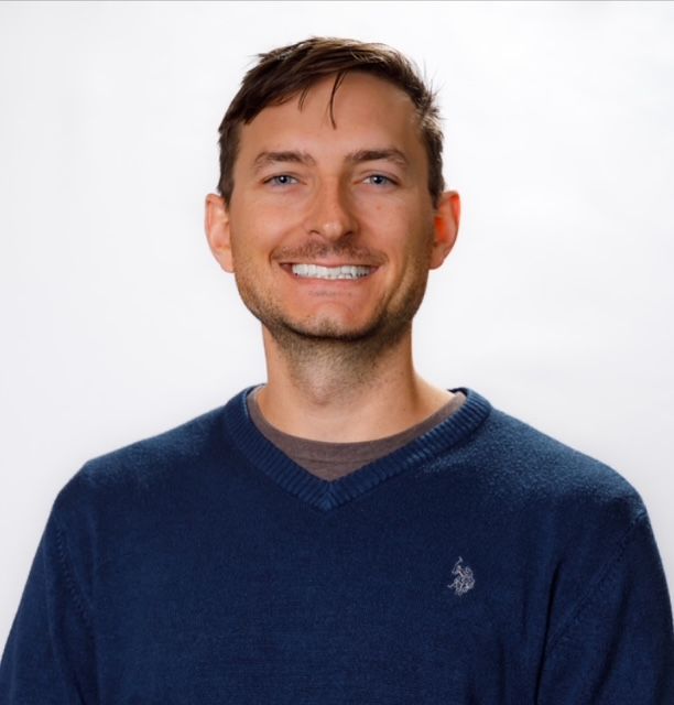
About Benjamin Colburn, DVM, DACVR
Practical veterinary radiology education grounded in clinical experience
I’m Dr. Benjamin Colburn, a board-certified veterinary radiologist at VitalRads in Cypress, Texas. My clinical work includes interpreting radiographs, ultrasound examinations, CT, and MRI studies, performing outpatient ultrasounds, and teaching veterinary residents.
I created DVMCE to help veterinarians develop a more systematic approach to diagnostic imaging and connect what they see on radiographs with practical next steps for their patients.
My clinical background
Working in diagnostic imaging means evaluating studies with a wide range of presentations ranging from routine examinations to cases that require urgent decisions.
My work involves identifying abnormalities, considering their significance in the context of the patient’s history, and communicating conclusions that help the treating veterinarian move forward with the case in the right way.
Through High Goal Veterinary Services, I also provide outpatient imaging at Foster Creek Veterinary Hospital in Richmond, Texas. Services include abdominal and thoracic ultrasound and ultrasound-guided fine-needle aspiration. For echocardiograms, I acquire the images and submit them to a board-certified veterinary cardiologist for interpretation and recommendations.
These experiences shape how I teach: start with a consistent search pattern, describe the findings clearly, and explain how those findings influence the next step.
Why I created DVMCE
I remember what it felt like to look at abdominal radiographs and wonder whether I was missing something important.
Is the gastrointestinal tract obstructed? Could the findings represent functional ileus? Is additional imaging needed? Is conservative treatment reasonable, or should surgery be considered?
I built my course around those questions. My goal is to make the radiologist’s reasoning easier to follow, using actual cases and clear explanations that you can bring back to clinical practice.
How I teach veterinary radiology
My teaching approach emphasizes:
-
A systematic search pattern: Develop a repeatable process for evaluating a study.
-
Normal anatomy alongside abnormal cases: Understand the reference point before interpreting the changes.
-
Short, focused lessons: Study a topic and return to it when you need a refresher.
-
Findings, conclusions, and next steps: Follow the reasoning from the image to the clinical discussion.
-
Honest recognition of uncertainty: Understand what the radiographs support, what they cannot exclude, and when further diagnostics may help.
I also participate in resident teaching and rounds. Explaining cases to other veterinarians reinforces the importance of clear descriptions and defensible interpretations, the same principles that guide my online education.
Is It Surgical? A Practical Guide to GI Radiology for the General Practitioner
My online course focuses on gastrointestinal radiology in dogs and cats. It walks through a systematic approach to the stomach, small intestine, and colon, with cases covering foreign bodies, mechanical obstruction, functional ileus, gastric dilation and volvulus, and other important GI disorders.
The course provides 2.5 hours of RACE-approved continuing education, with short lessons and one year of online access. It is designed for general practitioners, new graduates, and emergency veterinarians who want practical training in abdominal radiograph interpretation.
Education and credentials
-
Undergraduate education: Angelo State University, San Angelo, Texas
-
Doctor of Veterinary Medicine: Texas A&M College of Veterinary Medicine and Biomedical Sciences, College Station, Texas
-
Veterinary Diagnostic Imaging Residency: VitalRads, Cypress, Texas
-
Board certification: Diplomate of the American College of Veterinary Radiology — DACVR
Learn with me
If you want a clearer process for approaching canine and feline GI radiographs, I invite you to explore the course.